A new study investigates the transfer of information between neurons.
Do we really know how the brain works?
In recent decades, significant advancements have been made in comprehending the intricate workings of the brain. Researchers have gained extensive knowledge about the cellular neurobiology of the brain and have uncovered a lot about its neural networks and the elements constituting these connections. Despite this, a whole host of important questions remain unanswered and, consequently, the brain continues to be one of science’s great, tantalizing mysteries.
Perhaps one of the most nagging of these questions revolves around our understanding of the brain as a system. Scientists are still largely in the dark about how the brain functions as a network of interacting components, about how all the neural components cooperate, and especially, how information is processed between and among this complex network of neurons.
Revolutionary Research on a Simple Organism: The C. elegans Worm
Now, however, a team of neuroscientists and physicists at Princeton University are helping to shine a clarifying light on how information flows in the brain by studying, of all things, the brain of a very small but ubiquitous worm known as Caenorhabditis elegans. The details of the experiment are chronicled in a recent issue of Nature. The team consisted of Francesco Randi, Sophie Dvali, and Anuj Sharma and was led by Andrew Leifer, a neuroscientist and physicist.
“Brains are exciting and mysterious,” said Leifer. “Our team is interested in the question of how collections of neurons process information and generate action.”
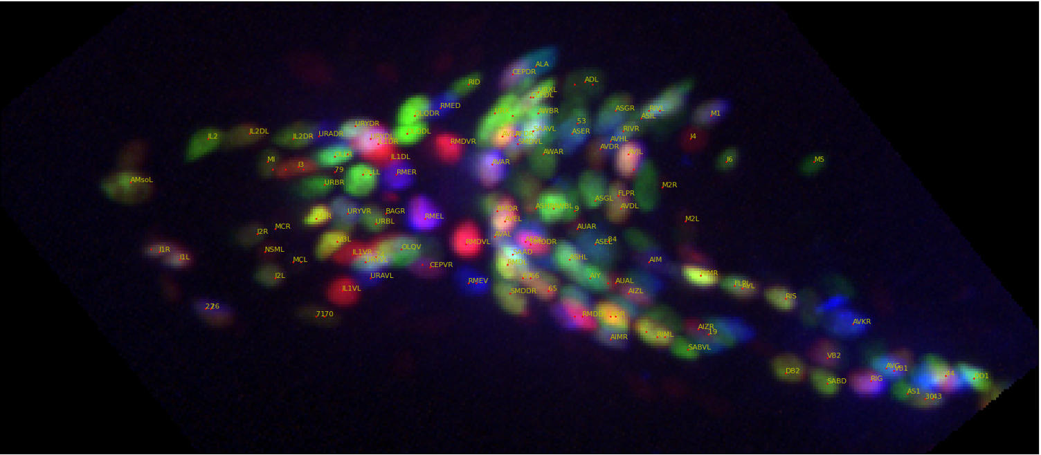
Video shows measurements of neural activity in the worm’s head as individual neurons are optically stimulated one at a time. The neuron in the crosshairs is stimulated when the words “Stimulated” appear. When neurons become active they appear dark red in this visualization. The video is sped up 4x. Credit: Francesco Randi, Princeton University
Interest in this question has broad implications, Leifer added. Understanding how a network of neurons works is a specific example of a broader class of questions in biological physics, namely, how collective phenomena emerge from networks of interacting cells and molecules. This area of research has implications for many topics relevant to biological physics as well as contemporary, cutting-edge technologies, such as artificial intelligence.
The first step in answering the question of how information is processed through a network of interacting neurons required that Leifer and his team find a suitable organism that could easily be manipulated in the lab. This turned out to be C. elegans, an unsegmented, non-parasitic nematode, or roundworm, that has been studied by scientists for decades and is considered a “genetically model organism.” Model organisms are commonly used in the laboratory to help scientists understand biological processes because their anatomy, genetics, and behaviors are well understood.
Innovative Techniques in Brain Mapping and Optogenetics
The worm is approximately one millimeter in length and is found in many bacteria-rich environments. Especially pertinent to the current study is the fact that the organism has a nervous system of only 302 neurons in its entire body, 188 of which reside in its brain.
“By contrast, a human brain has hundreds of billions of neurons,” said Leifer. “So, these worms are much simpler to study. In fact, these worms are excellent for experimentation because they strike just the right balance between simplicity and complexity.”
Importantly, added Leifer, C. elegans was the first organism to have its brain wiring fully “mapped.” This means that scientists have compiled a comprehensive diagram, or “map,” of all its neurons and synapses—the places where neurons physically connect and communicate with other neurons. This field of endeavor is called “connectomics,” in the parlance of neuroscience, and a diagram of a comprehensive map of neural connections in the brain of an organism is known as a “connectome.” One of the main goals of connectomics is finding out specific nerve connections responsible for particular behaviors.
An additional advantage in using C. elegans in laboratory experiments is that the worm is transparent, and, in certain cases, its tissue has been genetically engineered to be light-sensitive. This area of research is known as “optogenetics” and it has revolutionized many aspects of experimentation in biological neuroscience. Instead of the more conventional system of using an electrode to deliver a current into a neuron and thereby stimulate a response, the optogenetic technique involves using light-sensitive proteins from certain organisms and implanting those cells in another organism so that researchers can control an organism’s behavior or responses using light signals.
Similarly, other proteins can be used to light up and report when one neuron signals to another. This means two important things for laboratory experimentation: that an organism will respond to the presence of light, and that a neuron, once it receives a signal from another neuron, will “light up.” This has allowed researchers to study the interaction of neurons visually.
“What is really powerful about this tool is that you can literally turn neurons on and watch them signal in real-time,” said Leifer. “In essence, we can convert the problem of measuring and manipulating neural activity to one of collecting and delivering the right light to the right place at the right time.”
These optical tools allowed Leifer’s team to begin the painstaking task of understanding how information flows through the worm’s brain. The goal was to understand how signals flow directly through the worm’s entire brain, so each neuron had to be measured. This involved isolating one neuron at a time, shining a light on it, so that it was “activated,” and then observing how the other neurons responded.
“For this experiment, we went one neuron at a time through the entire brain, activating or perturbing each neuron and then watching the whole network respond,” said Leifer. “This way, we were able to map out how signals flowed through the network.”
“This was an approach that had never been done before at the scale of an entire brain,” added Leifer.
In all, Leifer and his team performed nearly 10,000 stimulus events by measuring over 23,000 pairs of neurons and their responses, a task that took seven years from conception to completion.
Challenging Established Models and Introducing New Insights
The research conducted by Leifer and his team is thus far the most comprehensive description of how signals flow through the brain. For scientists who study C. elegans, the researchers provided a lot of information on how specific signals work in the worm’s brain, and it is hoped that this research will provide a plethora of new information that will help advance basic research.
An equally important finding was that a number of the empirical observations Leifer and his team made during the experiment often contradicted the predictions of worm behavior based on mathematical models derived from the worm’s connectome map.
“We concluded that, in many cases, many molecular details that you can’t see from the wiring diagram are actually very important for predicting how the network should respond,” said Leifer.
The researchers suggest that there is a form of signaling—part of the “molecular details that you can’t see”—that does not progress along neural wires. Leifer and his group characterized these as “wireless signals.” Although wireless signaling is well known among neuroscientists, it has largely been underappreciated for studying neural dynamics because it had often thought to be a process that occurs very slowly. Wireless signaling is a form of signaling by which a neuron releases molecules, called neuropeptides, into the extracellular space, or “extracellular milieu,” between neurons. These chemicals diffuse and bind to other neurons even if there is no physical connection between them.
Finally, the researchers believe that an important impact of their work is that it allows other neuroscientists studying this and similar phenomena to develop better models with which to understand the brain as a system.
“With our research, we provided a very important piece of the puzzle that was missing,” said Leifer.
Reference: “Neural signal propagation atlas of Caenorhabditis elegans” by Francesco Randi, Anuj K. Sharma, Sophie Dvali and Andrew M. Leifer, 32 October 2023, Nature.
DOI: 10.1038/s41586-023-06683-4
This work was primarily supported by the National Institute of Health New Innovator Award, a National Science Foundation CAREER Award, and an award from the Simons Foundation. Funding was also received from an NSF Physics Frontier Center grant that supports Princeton University’s Center for Physics of Biological Function.
2 Comments
Your Doing studies on human subjects with these “c worms” and they have no idea. This is wrong and I’m not playing.
You’re using human subjects for your nefarious tests you put these worms in my eyes my brain I’m coming for you